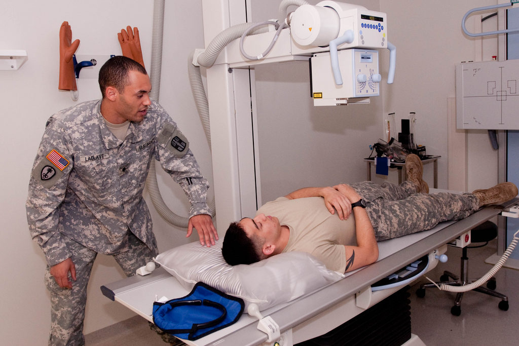
- Spc. Arbor L. LaClave, with the 322nd Medical Company Michigan Army Reserve Unit in South Field, Michigan works with his active duty counterpart, Spc. Justin J. Reichelt a radiology technician with the health clinic at Grafenwoehr Training Area. LaClave practice his spinal X-ray positions utilizing Reichelt as his mock patient to practice his skills as part of the Joint Multinational Training Commands overseas deployment training for reserve components.
There are many branches of medicine which are truly astonishing in terms of their history and the impact which they have had on various forms of treatment. Whilst not a doctor like my friend Apparao Mukkamala, I have long been interested in the various techniques which are used throughout medicine, how they came about and how they are still being shaped and improved in the modern world.
One such area that has always fascinated me is that of Radiology, a key medical tool which can be used for a wide range of treatments, assessments and diagnosis. If you find this type of thing interesting then I wanted to talk a little bit today about the history and origins of radiology, what it is, how it works and what the future looks like for this vital medical practice.
What Is It?
Radiology is the area of medicine which uses radiant energy that helps doctors to make diagnosis and that treats a wide variety of conditions and diseases. There are two key components of radiology which are diagnostic and interventional radiology. When used to make a diagnosis, the human body, or an area of it is exposed to radiant energy to create an image of what is going on below the surface. When used in treatment, radiant energy is blasted into a specific part of the body and can help to kill damaging cells.
A Brief History
Radiology came about when German physicist Wilhelm Conrad Röntgen made the discovery in his lab back in 1885, that an energized cathode ray tube could light up a wall covered with fluorescent material, from quite a distance away. The screen which was covered in the material was responding to the X-Rays which were being sent from the cathode and he began to tweak this discovery to blast images onto the screen. After being awarded the Nobel Prize in 1901, the German physicist went on to perfect his discovery and began using it on human bodies to study the skeleton.
Use
Through the years since its discovery, X-Rays and radiology have been used to diagnose and treat a wide number of ailments and there are many different facets to radiology. For example we use X-rays to look for broken bones in the skeleton, we can use MRIs (Magnetic Resonance Imaging) to take photos of various cross sections of the body and we can use Ultrasonography to listen to the inside of the body thanks to acoustic energy.
The Future
Radiography is still moving forward at an alarming rate and the improved use of technologies and computers is giving radiologists then chance to see deeper and more detailed images of the inside of the human body, as well as giving an incredibly detailed picture as to what is happening not just in the body, but inside cells and tissues throughout the human body. The key to beating many o the world’s diseases comes down to the speed of diagnosis and improved radiology will help medical professionals get to the bottom of problems in a faster manner.